
Tumour detection & 3D segmentation
Find it earlier,
measure it properly
Machine learning on routine MRI and CT to pick up brain tumours sooner, and to outline them on every slice as a true 3D volume rather than a single width.
Why a width isn't enough
A tumour is a
complex shape
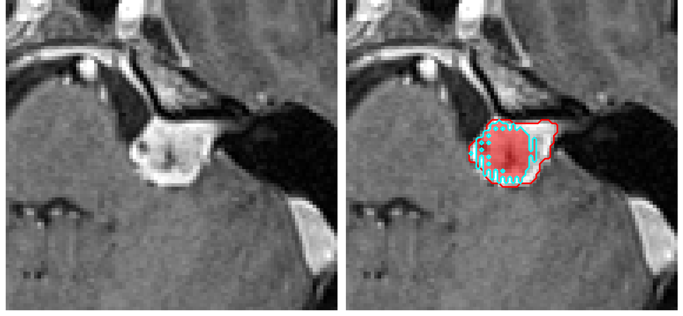
And it is unique in every patient. Report it by its widest diameter and you throw away most of what's there. Outline it on every slice and you get a real volume, and small changes show up long before they would shift a single measurement. The picture on the right is one of ours: the expert outline in red, our model's in cyan.
Finding it
Picking up brain tumours on routine MRI and CT, the kind that are easy to miss when they're still small.
Outlining it
Segmenting the whole lesion on every slice, automatically, instead of one hand-drawn diameter.
A real volume
A measurement in cubic millimetres, so a change of a few percent shows up long before it would move a width.
Cognia's imaging work is a research & development programme. It isn't a medical device and doesn't replace clinical judgement.
Request a demo
Twenty minutes is usually enough to work out whether we can help. Tell us about your service.